Osteoporosis advice at Seegarten Klinik
Osteoporosis is more than a lack of calcium: the bone loses mass and elasticity. In the osteoporosis consultation we assess your personal risk and show you how to strengthen your bones – with exercise, nutrients and, where necessary, further therapies.
Osteoporosis is rarely a disease of the bone; it is usually a reflection of systemic disorders.
The consultation is intended for women and men with a diagnosis of osteoporosis, with risk factors, or with questions about an ongoing treatment.
What bone density measurement tells us – and what it does not
Today the diagnosis of osteoporosis is usually based on bone density measurement (DEXA) and the T-score calculated from it: if it is below −2.5, the bone is considered osteoporotic.
The measurement has its limits. Density is mass per volume; DEXA, however, scans the bone in only two dimensions and measures an areal density in grams per square centimetre. Devices, reference databases and correction factors differ, and the measurement itself varies by up to 3% at the lumbar spine and up to 5% at the hip.
What DEXA does not show
- the strength of the bone
- its structure and quality
- its ability to repair itself
- its metabolism
Bone density also says little about the risk of fracture: the increase in bone density explains only 4 to 28% of the reduction in fractures (Li 2001, Cummings 2001, Sarkar 2001).
The T-score does not know your weight
The T-score compares your bone density with that of healthy adults aged about 25 to 30. Disease is thus defined as distance from youth. Age, sex and height go into the evaluation – body weight does not.
But bones carry weight: the heavier a person, the denser their bones. Many slim women are therefore given a diagnosis of osteoporosis although they simply have light bones for a low weight. The same is true of the muscles.
Anyone in the yellow or red area but above, on or just below the line needs no treatment. In the usual evaluation, all these people are considered osteopenic or osteoporotic – and are given an osteoporosis therapy they do not need at all.
Conversely, an overweight person can have an osteoporotic constellation despite normal bone density – namely when their B-score is low. This is rare, however.
The B-score: bone density in relation to weight
The B-score (“bone score”) relates your measured bone density to your body weight. The normal range is between 0.87 and 1.13; below that, bone density is low for your weight. Enter the bone density of the lumbar spine (L1–L4) from your DEXA report and your weight.
For research purposes only: the B-score is not a recognised diagnostic criterion and does not replace a medical assessment. Your entries and the result are stored on our server without your name and evaluated statistically. © 2024 John van Limburg Stirum
Please enter the year of the DEXA measurement for the printout.
Please bring the printout with you to an appointment at the SGK.
How we investigate
We look for the causes. Osteoporosis can be the consequence of other disorders:
- Hormonal: overactive parathyroid glands, overactive thyroid, Cushing's syndrome, lack of sex hormones
- Inflammatory: rheumatoid arthritis, chronic inflammatory bowel disease, chronic kidney failure
- Blood and tumours: multiple myeloma
- Neurological: immobility, Parkinson's disease, after a stroke
- Functional: loss of muscle (sarcopenia)
Medication can also weaken the bone:
- Cortisone
- Antidepressants
- Stomach protectants (proton pump inhibitors)
- GnRH analogues
- Aromatase inhibitors
- Antidiabetic drugs (glitazones)
- Anti-epileptic drugs
- High-dose thyroid hormones
Laboratory tests in osteoporosis
Laboratory tests can follow what the density measurement does not show – the metabolism of the bone:
- Bone breakdown: CrossLaps in the blood, deoxypyridinoline in the urine
- Bone formation: procollagen type 1 (P1NP) in the blood
- New: calcium isotopes in the blood – the ratio of formation to breakdown
Find out more: Bluttuning, preparing for the laboratory.
How we treat
The problem in osteoporosis is a loss of bone elasticity (because the calcium salts predominate) and of bone mass (because the organic bone substance, the proteins, is broken down more quickly). Under these conditions, accidents with awkward falls in old age are more likely to result in a broken bone. In advanced osteoporosis, however, even minor “traumas” such as lifting a shopping bag, bending down or merely sneezing can cause a fracture.
Preventing fractures
A broken bone rarely has just one cause. Besides osteoporosis, what counts is the loss of mobility and balance, declining eyesight and reaction, loss of muscle – and the risks a person takes. This is where we start:
- Improving mobility: exercise, hopping
- Strength training
- Enough protein – which includes good teeth and vigorous chewing
- Improving balance
- Optimising eyesight
- Training the ability to react
- Dietary supplements – but no calcium
The fact is that age-related lack of exercise, with muscle weakness and an unsteady gait, is the most common cause of fractures, which is why therapeutic measures should begin precisely here. Regular physical activity with light strength training not only builds up the muscles, but also improves mobility, agility and the ability to avoid dangerous situations.
From a medical point of view, it is precisely older people who can and should significantly increase their quality of life, their sense of well-being and their life expectancy through activity. Studies from the USA recorded an increase in strength of more than 50% (!) in people over 80.
Sport: bone needs loading
Mechanical loading is the decisive stimulus for building bone; without loading, bone is lost quickly. What matters is the force acting on the bone:
Acid-base balance
An alkaline environment activates the bone-building cells (osteoblasts) and promotes mineralisation. Acid inhibits formation and promotes breakdown. Balancing the bases is therefore part of the treatment.
Supporting new bone formation with nutrients
- Vitamin K (new bone formation)
- Vitamin D (incorporation of calcium)
- Magnesium (activation of vitamin D)
- Manganese (needed for the connective tissue of bone and for mineralisation)
- Folic acid (prevents the formation of homocysteine, which harms the bones)
- Vitamin B6 (reduces homocysteine, cross-linking of collagen fibres)
- Vitamin B12
- Vitamin C (osteoporosis in vitamin C deficiency, i.e. scurvy)
- Boron (vitamin D metabolism)
- Silicon (stabilises collagen through cross-links)
- Zinc (increases the activity of vitamin D)
- Copper (cross-linking of collagen fibres)
- Molybdenum
- Potassium citrate (base balance)
- Isoflavone (a plant compound that binds to the oestrogen receptors of the bone cells)
Enough protein and collagen are part of this. From about the age of 55, or when taking stomach protectants, stomach acid is often lacking; protein and calcium are then absorbed less well.
Calcium: not as a tablet
More lime does not mean more bone. Calcium tablets, however, carry risks: in a meta-analysis, pure calcium preparations increased the risk of heart attack by up to 30 per cent, especially at more than 800 milligrams a day (Bolland et al., British Medical Journal 2010). The German Society of Endocrinology states: “Calcium intake by tablet, if dosed above 500 mg/day, can result in kidney stones and vascular calcification” (2022).
Medication
Osteoporosis drugs work in two ways: they inhibit bone breakdown (bisphosphonates, denosumab, raloxifene) or stimulate formation (teriparatide, romosozumab). Whether a drug makes sense for you is something we discuss in the consultation – together with what you can do yourself.
Find out more: Sports Medicine for strength training, Endocrinology & Diabetology for hormones. In the MediWiki (in German): Vitamin D, acid-base balance.
A patient's case
This patient was 61 when she was faced with a diagnosis of osteoporosis. She was subsequently given bisphosphonates, drugs that inhibit bone breakdown. By 2009 a first success could be recorded on the basis of the bone density measurement. Because of side effects, she was switched to a preparation containing calcium and vitamin D. Two years later a decline in bone was recorded again. For this reason we changed the treatment to our special micronutrient therapy (free of side effects). It can be seen quite clearly how bone density has increased steadily since then. Bone density was measured again recently. The patient is now 73 (!) and showed a further increase in bone density of 15% within the last three years! The radiologist's comment: “I can't explain that.”
We can.
Background: the osteoporosis story
What osteoporosis is – and what it is not
Osteoporosis is a specific disease that must be clearly distinguished from the normal decrease in bone density in old age (so-called osteopenia). In the past, bones were described as osteoporotic only once fractures had occurred – usually of the spine, the hip or the wrist – as a result of disproportionately minor trauma. According to the newer definition, any decrease in bone mass is now osteoporosis, even if there are no fractures. With the ageing of the population, and with the lack of exercise and poor nutrition that come with modern life, osteoporosis is nevertheless becoming more and more apparent. Of course, this condition already existed in earlier times.
The “witches” who appear in history as old, stooped women confirm this observation. How many women were burnt as witches merely because they suffered from osteoporosis must remain an open question.
However widespread and well researched this change in the bones may be, there are major contradictions in the definition of the disease and therefore also in the recommended therapeutic measures. A selection follows:
In medical school, every student had to know precisely the difference between osteoporosis and so-called osteomalacia. The former consisted in a “wasting” of bone, the latter in a softening of bone. A wrong answer in the examination would have cost a good many marks. Nevertheless, clinical, pharmacological medicine practically equates osteoporosis with osteomalacia. Wherever the cause and treatment of osteoporosis are discussed, calcium deficiency is cited – and that is precisely what causes osteomalacia! In the student textbook of orthopaedics by Dr Alfred M. Debrunner, the development of bone in old age is (correctly) explained as follows: “While in the bone of a child the proportion of organic substances (collagen) is still relatively large and the bone is still flexible, in old age the inorganic proportion (calcium salts) increases steadily, and the bones become more rigid and more brittle.” According to the WHO, “osteoporosis is a disease characterised by low bone mass and deterioration of the fine structure of bone tissue, leading to increased bone fragility and ultimately to a higher risk of fracture”. There is no mention of calcium in the WHO documents either. We can therefore see at once that osteoporosis is not a calcium problem and is not solved by giving calcium.
Why calcium alone does not help
That bone formation in no way depends on “topping up” with calcium is shown by the following phenomenon: a cow gives 20–25 litres of milk twice a day. This contains on average 1% lime, which corresponds to a daily calcium loss of 400 g. Yet, as a strict vegetarian, the cow consumes none of the “official” sources of calcium recommended for humans. This raises the awkward question: where does she get the lime from? The cow suffers neither from calcium deficiency nor from osteoporosis, although she gives the milk and does not drink it!
So what happens when the organism is offered calcium in “sufficient quantity”, as usual with vitamin D? The organism is then forced to incorporate the calcium. But since calcium can only be incorporated into an organic substance that is already there, and there is not enough of it in the bone, it has to be deposited somewhere else – for example in arteriosclerotic plaques of the aorta. Indeed, one comes across X-rays in which the calcium-laden aorta appears denser than the spine.
A lack of lime in childhood is rickets. Nevertheless, supplying lime has no effect.
What bone density measurement measures
A further discrepancy arose with the introduction of bone density measurement (so-called densitometry). This procedure is supposed to detect osteoporosis and assess the success of a therapy. But once again the method misses the point. What interests us is the risk of a pathological fracture. Resistance to fracture, however, does not depend on the calcium content (and that is the only thing densitometry measures), but on elasticity. What we want in old age is youthful bones, “children's bones”. They are almost impossible to break.
Why? Because they are flexible and not yet calcified! If you put a child's bone into the densitometer, however, the result would inevitably be severe osteoporosis! The bones can hardly be made out. And of course this method can be used to “prove” that calcium therapy combats osteoporosis. Depositing lime merely simulates new bone formation, by calcifying trabeculae that were previously invisible on X-ray (because still cartilaginous and uncalcified). For fun, place a column of lime such as a stick of chalk in the X-ray beam and you will get a fantastic bone density. But … there is hardly anything more brittle!
Summary report of the working committee “Medical Treatment” of the Federal Committee of Physicians and Health Insurance Funds on its deliberations in 1999 on the assessment of osteodensitometry under §135 para. 1 SGB V, 22 March 2000
Osteodensitometry is widely used nationally and internationally to diagnose osteoporosis, but it has been medically controversial for years. Since osteodensitometry was developed and came into use, expert opinions and statements have repeatedly been published in the scientific literature, at home and abroad, that call into question the validity and the benefit of this diagnostic method in part or even as a whole. It is known, for instance, that only some patients with increased bone fragility show low values in osteodensitometry, and conversely many patients with low values suffer no fractures. In other words, the relationship between the value measured by osteodensitometry and osteoporosis-related fractures that actually occur is not reliable. What is doubted above all is the use of the method to decide on preventive measures that have to be continued for many years, up to several decades. These include, for example, hormone replacement in women after the menopause, or the administration over years of other highly potent drugs that affect bone metabolism.
Oestrogens
It is well known that they suffer from osteoporosis more often than men. And where is the little difference? Quite right! Women produce oestrogens and men do not. As long as women have oestrogens, they are said to be protected against osteoporosis. After the menopause, things go downhill. The entirely natural female postmenopause (in Japan there is not even a word for the menopause) has been turned by Western medicine and the press into an oestrogen deficiency disease, and is a further expression of today's reductionism in medicine. Oestrogens are called for. But here we meet further contradictions. Bone loss has been found in women with so-called anovulatory cycles, which means that ovulation does not occur. In these women, however, it is not oestrogen that is lacking, but progesterone. Indeed, it has already been reported from the USA that giving progesterone alone increases bone mass. Oestrogens, at most, have a revitalising effect.
Oestrogens would have to be taken for about 20 years in order to maintain high bone density into old age. This medication is not free of side effects, however, since the risk of cancer of the womb or of the breast can rise. Nobody knows today whether the benefit really outweighs the harm.
Overweight women are fortunate. They produce oestrogens in their fatty tissue and are therefore immune to osteoporosis (according to the prevailing view in conventional medicine). Here again an explanation is attempted at the lowest level of medical knowledge. The fact is that osteoporosis is a very complex process with a still unmanageable number of unknown factors. But simple observation and trust in nature may help us here. At the latest since space travel it has been known that weightlessness can very quickly trigger osteoporosis. Mother Nature says to herself: why strong bones if I have nothing to carry? That is also why many slim patients show “osteoporosis” in the bone density measurement. Here they become victims of statistics (the person of normal weight is heavier). Overweight people depend on heavy, denser, stronger bones and for this reason show a “lower” risk of osteoporosis. In return, their bones also have to absorb more weight in a fall.
Osteoporosis campaigns
Again and again we are confronted with such campaigns. They are unmistakable sales instruments for drugs from the pharmaceutical industry. Approaches such as sport and nutrition are mentioned, but drug treatment always remains in the foreground, as a rule with the so-called bisphosphonates (a business worth billions, so-called “blockbusters”). These prevent bone breakdown, but in doing so they also cause the bone substance to become over-aged. Moreover, taking such medicines must not lull one into a false sense of security: “Nothing can happen to me now …”. Nutrition and sport, especially anti-gravity training as in a fitness club, remain the BEST conditions for bone health over many years.
So … at the next campaign, do not let yourself be talked into drug therapy too quickly. Always discuss the advantages and disadvantages, and especially the alternatives, with your family doctor.
Despite the many open questions and contradictions, it is possible for each individual to estimate their personal risk of osteoporosis and – with some motivation, starting as early as possible and persevering for years – to protect themselves against it.
“Puls” of 26 February 2018: problems with Prolia
Watch the programme on SRF (in German)
Comment by Dr. med. John van Limburg Stirum
The programme speaks of a “rebound effect”. It is surprising that the scientists are astonished by this phenomenon. It is known of all biological systems that any suppression of a natural function is afterwards answered with a counter-reaction. Walking barefoot in the snow is one example: afterwards the feet are hot! This phenomenon does not occur, however, when poisoning is involved, as with the bisphosphonates that inhibit bone breakdown. The recommendation of conventional medicine to taper off the therapy with bisphosphonates after using these monoclonal antibodies is therefore remarkable. In other words: first suppression, then poisoning.
Fortunately, there are natural remedies and procedures which, by contrast, promote the building of bone and may thus be an alternative to the standard therapy.
Advertisement for an osteoporosis drug – August 2019
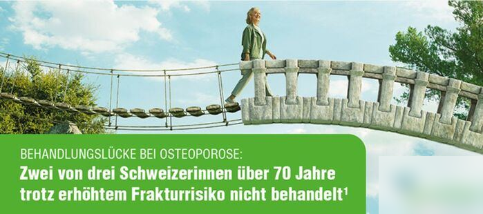
A provocative question …
Which bridge would better survive an earthquake?