Blood pressure, blood lipids and circulation are linked to metabolism, inflammation and lifestyle. We investigate before symptoms arise and support you once they are there.
The following sections present the procedures.
Vascular chelation therapy
Arteriosclerosis (calcification) of the abdominal aorta
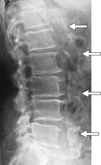
Chelation therapy is an outpatient treatment that is currently enjoying a real triumphal march through the USA. Many books and scientific publications document its outstanding results in arteriosclerosis and circulatory disorders. Among other things, the body is given a solution that contains various minerals and vitamins, but above all the active substance EDTA (see below). A similar compound also occurs in protein-rich foods.
EDTA has the property of binding heavy metals in the body, for example cadmium (cigarettes) and lead (petrol residues), as well as the light metal calcium, and excreting them via the kidneys. It is therefore outstandingly suitable both for metal poisoning and for the treatment of arteriosclerosis. The decisive point is that EDTA has a great binding capacity for the so-called transition metals, which can be helpers in the formation of excess free radicals. As a result, arteriosclerosis is halted, the cell functions are restored and the calcium overload is reduced. Calcium bound to protein is left untouched by EDTA. No decalcification of the bones occurs.
Chelation therapy is practised all over the world. In the USA, more than 1000 doctors have already specialised in it. Around 300,000 patients have so far received approx. 4,000,000 infusions.
The history of chelation therapy began in the 1930s at the company IG Farben in Germany, where a white, crystalline, water-soluble powder of ethylenediaminetetraacetic acid (EDTA) was developed as a colour stabiliser. A few decades later, the usefulness of this powder for medicine was also discovered. In the 1950s, an American doctor who suffered from very severe angina pectoris came upon the idea, in his mortal distress, of decalcifying his arteries with EDTA. This saved not only his own life but also the lives of many of his heart patients, whom he called in to be treated with his new method. In the years that followed, chelation therapy was used by a constantly growing number of doctors.
The effect of EDTA
- EDTA is a synthetic amino acid that is non-toxic to humans and has the ability to form complexes with metal ions (= “chelates”: from the Greek chele = claw), from which these cannot escape, like “prisoners in handcuffs”, and are transported out of the body (decorporated) with the urine. The first uses of chelation therapy were therefore in acute poisoning with the heavy metal ions lead, cadmium and mercury. Today it is known, in contrast to the knowledge of the chelation pioneers, that the effect of EDTA cannot be compared with a coffee machine descaler but is extraordinarily complex:
- EDTA increases the formation of parathyroid hormone in the parathyroid gland. This supports the removal of calcium from deposits in unphysiological places (arteries) and takes it to the places where it belongs (bones and teeth). This is the basis of the slight recalcification of osteoporotic bones under therapy with EDTA.
- EDTA acts as a potent antioxidant against the cell-damaging effect of the so-called free radicals by removing the corresponding excess catalysts (effect enhancers) copper and iron from the body.
- EDTA eliminates the heavy metals lead, mercury, cadmium and aluminium and thereby restores the proper function of the enzyme chains.
- EDTA stabilises the cell membranes.
- EDTA ensures balance in the prostaglandin hormone system and free blood flow to the individual organs.
- EDTA lowers the risk of the formation of a thrombus, which blocks the coronary arteries in a heart attack.
- EDTA increases the elasticity of tissues, improves skin tone and reduces the formation of wrinkles.
The scientific foundations
for today’s practice of chelation therapy were also provided by other branches of medicine such as nutritional science, orthomolecular medicine, enzyme therapy, cancer and rheumatism research, as well as the physiology and biochemistry of metabolism and of oxygen supply in chronic, degenerative diseases. All these research results led to the discovery that the common cause of the degenerative diseases and of the ageing processes is not only the heavy metals and the calcium and cholesterol waste deposits on which the first chelation therapists focused their attention, but that the roots of all these diseases lie “one storey” deeper, with the so-called free radicals. When everything works well, these supply, as “energetic projectiles”, the energy needed for metabolism; but as soon as their capture and transfer mechanisms are defective, they damage the body’s own tissue cells by “bombarding” its own cells right down to the chromosomes, which are responsible for cell division, and cause them to multiply in an uncontrolled way, which is the cause of all malignant tumours. Chelation therapy and the reduction of free radicals enable the damaged cells to recover slowly.
| Main indications | Secondary indications |
|---|---|
| Arteriosclerosis Angina pectoris Rheumatoid arthritis Diabetes Vascular occlusions Skin conditions Cardiac arrhythmias Detoxification for smokers | Alzheimer’s disease Eye conditions such as macular degeneration Frigidity and impotence Multiple sclerosis Kidney calcification Rheumatism Shoulder tendinitis Scleroderma |
Various study results
from: Altern. Med. Rev. 2007 Jun; 12(2):152-8
EDTA Cardiac Events Study: 220 patients with 3 years of follow-up
| Statistically predicted number | Actually occurred | |
|---|---|---|
| Heart attacks | 15 | 0 |
| Deaths | 6 | 0 |
| PTCAs during follow-up | 31 | 2 |
| CABG operations during follow-up | 16 | 6 |
Surgical or interventional procedures required in patients with primary PTCA and stent therapy during the 3-year follow-up
| Control groups | Patients treated with EDTA (25 patients) | |
|---|---|---|
| Re-PTCA | 22.3% | 4% |
| CABG operation required | 11.8% | 0% |
Thrombosis of drug-eluting stents with clopidogrel compared with EDTA
| EDTA | Long-term administration of clopidogrel | |
|---|---|---|
| FDA approval | No | No |
| Clinical studies | No | No |
| Platelet inhibition | 3 mechanisms | 1 mechanism |
| Risk of major bleeding | No | Yes |
| Preliminary clinical data showing efficacy | Yes | Yes |
Study by Dr. Thomas B. Fischer
- Thinning of the inner vessel wall after 30 chelation therapies
- IM T re / li = measurements before therapy
- re / li n. = measurements after therapy
- 0 = average values
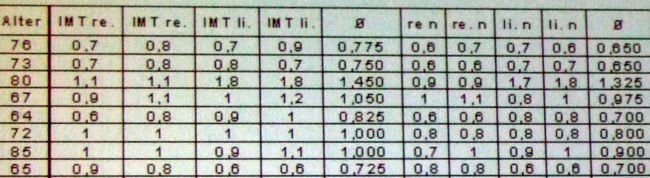
The results are highly significant.
Today’s chelation therapy, with its vitamin and mineral additives, has thus become not only a revolutionary causal therapy for chronic degenerative diseases that already exist but also, when used repeatedly as a preventive measure, a preventive therapy for the ageing processes and the complaints of old age as such, whose appearance it can postpone by years.
The insufficient spread of chelation therapy
and the medical profession’s lack of knowledge about this method can be explained by the fact that the patent for EDTA expired in 1948 and EDTA is not a lucrative product from a commercial point of view, so that the pharmaceutical companies are not motivated to advertise the unique effect of chelation therapy. Heart surgeons and specialists in conventional medicine also lack the motivation to learn this method, although they are obliged by law to inform the patient about the alternatives to a heart operation. According to statistics from the USA, up to 60% of bypass operations could be avoided by chelation therapy.
Costs
The costs of the therapy can be found in the current price list (under login) and include the regular urine and blood pressure checks and all additives (vitamins, trace elements and others) to the infusion. The price results above all from the duration of several hours and the monitoring of the infusion. The total costs thus depend on the severity of the illness and the corresponding number of infusions required.
Contraindications
The contraindications for treatment with the EDTA solution are insufficient kidney, liver and heart function, pregnancy and calcified zones of tuberculosis. As EDTA binds not only calcium but also essential trace elements and lowers the blood level of vitamins B and C, these must be added to the chelation infusion (substituted). For this reason, medical supervision before and during chelation therapy is necessary.
Side effects
Occasionally, slight pain, tiredness and a change in blood pressure or blood sugar level may occur during the infusion. They are controlled without difficulty by slowing the infusion rate. These accompanying symptoms usually disappear by themselves after 2 sessions.
As other minerals such as calcium, iron, manganese, molybdenum and zinc may also be excreted, these must be checked regularly (approx. every 10 infusions).
Preparation
A thorough medical examination (with blood and urine analyses), also periodically during the treatment, ensures optimal monitoring.
The treatment
A complete course of chelation therapy comprises a minimum of 20 to 30 individual infusions, a healthy diet and sufficient exercise. In advanced cases of arteriosclerosis and in arterial occlusion, considerably more sessions are necessary (30 to 100 infusions); as a basic rule, the number of infusions required corresponds to the patient’s age. The infusions are given 2 to 3 times a week and must be administered very slowly. A therapy session therefore takes at least 3 to 4 hours. A burning sensation during the therapy from the puncture site upwards means that the infusion rate must be reduced. To strengthen the effect of chelation therapy, additions of vitamins and trace elements and booster infusions at regular intervals are recommended.
Chelation therapy can also be used together with other forms of therapy for arterial occlusive diseases. It is entirely compatible with “blood thinners” and with medicines that dilate the blood vessels, lower blood pressure or act against cardiac arrhythmias. During or after successful chelation therapy, it is often possible to do without certain medicines in part or entirely. Chelation therapy is also possible after vascular surgery or amputations. The aim, however, is to use this therapy to prevent such serious interventions from the outset.
Left
Report from the EUROPÄISCHE ZEITUNG, March 2001
Not recognised by conventional medicine despite demonstrable successes
Chelation therapy slows “free radicals”
When the term chelation therapy is mentioned in medical circles, an extremely controversial discussion can be expected with certainty. This is because conventional doctors have always taken a critical view of new therapeutic principles and it takes a long time for the “new” to become socially acceptable.
The American sailors were actually only supposed to paint their warship. But this task had dangerous consequences: during their work, the seamen inhaled the toxic fumes of the lead-containing red lead paint. This was a decisive test for the new chelation therapy. With the help of a chelating agent, the so-called EDTA solution, the sailors’ lead poisoning was treated, and successfully in every case. That was not all: it was soon found that in some patients further complaints, above all anginal symptoms, also disappeared and that people’s general condition improved markedly.
That was in the 1950s. The positive effects of chelation therapy that had first been found in the sailors could later also be observed in a similar form in children who had scratched lead-containing wallpaper off the wall and put it in their mouths.
The binding capacity of EDTA (Ethylene-Diamine-Tetraacetic-Acid) for heavy metal ions was known. The substance itself dates from the 1930s, when it was used in the textile industry. The principle of chelate compounds goes back to the Swiss Nobel laureate of 1913, Alfred Werner.
It was first used in humans as early as the war years and afterwards, to treat radioactive contamination and nickel poisoning.
Between 1950 and 1966, chelation therapy experienced a “heyday” in the USA, although at that time, measured against today’s state of knowledge, there were still no clear ideas at all about the dosage and the infusion time. At the end of the 1960s, the rise of bypass operations began in the USA, and chelation therapy largely disappeared from the scene. In addition, there were some deaths under chelation therapy, which were published and understandably spread uncertainty.
These deaths, however, can clearly be attributed to errors in dosage and infusion time: from today’s point of view, about twelve times the amount of EDTA was administered at that time, and the duration of the infusion, today three and a half to four hours, was about half an hour. The patients developed a crash kidney and died of acute kidney failure.
All of this now lies more than 30 years in the past, and in the meantime there is absolute clarity about the correct dosage and duration of infusion. Nevertheless, these incidents are still cited today when it comes to discrediting and rejecting this therapy.
What is studiously overlooked, by contrast, is that according to reliable statistical data not only do around two to three per cent of patients die during or after bypass operations, but about 50 per cent of the bypasses close again after a few years. This information is not usually passed on to patients in the relevant information discussions.
The approach of a causal therapy
But what lies behind chelation therapy? And above all: how does it work? Every person has vessels in their body with a length of about 100,000 kilometres, including the capillaries. In a bypass operation, a stent or a balloon dilatation, the corresponding measures are carried out over a length of a few centimetres, and the “rest” remains untreated, so one can certainly speak of symptomatic measures. In chelation therapy, the infusion fluid (EDTA solution) is distributed throughout the entire vascular system: the approach to a causal therapy.
While the binding of heavy metal ions by the chelate in the course of chelation and their excretion (over 90 per cent via the kidneys) have long been known, until a few years ago there was still the idea that chelation therapy was a kind of “drain cleaner” for the calcified vessels. EDTA was thought to loosen the arteriosclerotic plaques, which would then be flushed out by the blood. This raised the obvious question of whether larger plaques might not then lead to infarctions.
Pharmacological investigations have shown, however, that the mechanism of action of chelation therapy is essentially to be seen in a minimisation of free radicals. These free radicals are recognised as one of the main causes of arteriosclerotic processes. The pathology of free radicals is a cornerstone of today’s knowledge about the mechanism of action of chelation therapy.
The main indication for this therapy is vascular disease caused by arteriosclerosis.
The following should be mentioned in particular:
- Intermittent claudication (“window-shopping disease”)
- CHD (coronary heart disease)
- Condition before recommended bypass operations (if not so acute that surgery must be performed immediately)
- Condition after bypass operations (to keep the vessels open permanently)
- Condition after apoplexies (strokes) and infarctions
- Arterial occlusions and stenoses in general
In addition, the elimination of heavy metal ions remains an important indication, and very often the amalgam burden can lead to considerable health problems.
Provided that the requirements of the therapy protocol issued by the DGCT (Deutsche Gesellschaft für Chelat-Therapie), which is structured in line with the therapy recommendations of the American ACAM, are followed in chelation therapy, experience shows that only minor side effects are to be expected. The most frequent side effects are burning at the puncture site, shivering (temporary), a fall in blood pressure and tiredness.
Of course, the positive “side effects” that can frequently be observed should not be disregarded either: lowering of total cholesterol, improvement of vascular tinnitus, improvement of vision, gradual lowering of blood pressure (reduction of antihypertensive medication), improvement of anginal complaints.
In general, patients with severe heart disease, with severe kidney and liver disease and, naturally as a matter of principle, pregnant women should not be treated with chelation therapy.
As chelation therapy, as already mentioned, is not yet recognised by conventional medicine, the costs are not reimbursed by the health insurers, which means that the patient has to pay for the therapy personally (exceptions in cases of heavy metal poisoning).
Chelation therapy is already being used preventively to an increasing extent, on the one hand out of fear of events that run in the family (apoplexies, infarctions), on the other hand as a contribution to securing future health, for greater resilience and thus also for stronger professional and private creativity.
Michael Brückner
Patient comments
Details of how I feel as a result of chelation therapy (16 treatments)
- My blood pressure has stabilised at the normal level, so that the blood pressure medication could be discontinued.
- Stabbing pains in the neck region have disappeared.
- I feel more energetic, more focused.
- Tightness and feelings of anxiety, above all at night and during physical exertion (rushing for the train), have disappeared.
So I am very grateful and happy about this good progress and would like to thank you warmly once again for everything.
Mrs R.J.
CT angiography check: in the area of the internal carotid artery (the artery in the neck), stenoses (narrowings) are no longer detectable on either side.
***
I now have 36 chelation infusions behind me and can report the following. I now often have to switch off my hearing aid because it is really too loud for me, so I hear better. Before the therapy I had the feeling that I needed stronger spectacle lenses. In the meantime I have new glasses with the same lenses for distance. Only for reading have they become a little stronger. So I see better. Now comes my greatest joy: my memory. I walk through the village again and greet people by name. I go down to the cellar and still know what I wanted to fetch. Before, it was often depressing. I am very, very grateful for this success.
M.M., aged 61, 3 October 2006
***
After only 5 treatments I felt an increase in circulation. Suddenly I could run better without developing angina pectoris. The night-time calf cramps have disappeared. Breathing is easier for me. The blood flow to my head is better too. My eyesight when reading has also improved. Disappearance of unpleasant symptoms of Lyme disease such as joint and nerve pain. My memory has improved and my joy in life has returned.
I.A., aged 65, 17 November 2006
***
Since the chelation therapy I feel warmer in general and can tolerate the cold much better.
U.S., aged 60
***
Chelation therapy has given me something like a second life. No more back pain, and my dizziness has disappeared completely. My general well-being has improved and my resilience has increased. My cardiac arrhythmias have almost disappeared.
H.B., aged 86
***


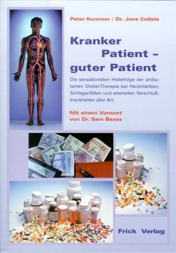
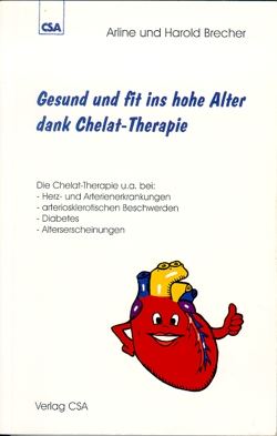
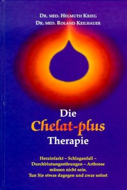
Recommended books


Bloodletting
Here, as a rule, approx. 200 ml of blood is taken from the vein of the arm. Bloodletting serves to relieve the circulation, to purify the body and for elimination.
If one has blood that is too “thick”, with a disturbance of the flow rate, the supply of oxygen and nutrients and the removal of harmful substances and metabolic products may be slowed down. Certainly, blood withdrawals were also abused in the past, which brought them into disrepute; GALEN and HIPPOCRATES made masterly use of them, while many physicians, above all French ones, claimed victims through carelessness, negligence and exaggeration and often led to the rejection of such a “dangerous” method.
Only today, now that blood withdrawal is coming back “from America” for the prevention of heart attacks, or leeches are becoming acceptable again in reconstructive and plastic surgery, is conventional medicine realising that there must be “something” in it. In impending pulmonary oedema, in eclampsia, in impending uraemia and in polycythaemia vera, conventional medicine “permits” bloodletting, i.e. one is at least not accused of malpractice for using it, one is merely smiled at. Yet the range of indications for blood withdrawals is so broad that many possibilities and opportunities are wasted in internal medicine, in traumatology and in gynaecology if blood withdrawals are not used.
What happens during bloodletting?
Because a “portion” of blood is removed, new blood has to be formed. These young blood cells are more elastic than the “old” ones. As a result, the overall fluidity of the blood increases slightly. However, because the same blood circulates thousands of times in the circulation, this small effect is “multiplied” and results in substantially improved blood flow in the longer term.
The fact that women remain protected from arteriosclerosis and heart attack until the menopause is probably due, among other things, to their physiological monthly “bloodlettings”.
Procedure
Until the desired haematocrit (the proportion of red blood cells in whole blood) is reached, which is around 42% in men and around 40% in women, portions of 200 - 250 ml are as a rule taken every 2 weeks. It is important to drink a lot, a great deal, before the bloodletting and on the same day: at least 2-3 litres of water.
Heart rate variability
- How adaptable is your heart to internal and external stimuli?
- Is your heart constantly on high alert?
- Can your heart recover?
- What is the ratio between tension and relaxation?
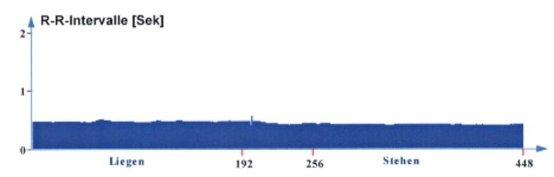
Measuring heart rate variability (HRV) is the first step towards a new balance. It records the heart rhythm lying down and standing. From this, the activity of the autonomic nervous system (sympathetic and parasympathetic) and the health of the heart and circulation are derived.
How do I know when I am stressed?
Sometimes you notice quite clearly that you are on high alert, for example when your pulse is raised. But people do not always perceive a stressful situation as such. Anyone who wants to take a close look at their stress profile in everyday life should have an HRV measurement carried out.
Heart rate variability indicates the time intervals at which our heart beats and is therefore a measure of the heart's capacity for regulation. The more the heartbeat varies, the better the heart adapts to the environment and to internal stimuli (thoughts, feelings, illnesses). The reverse also applies: the more regularly the heart beats, the more stress the body is experiencing.
Heart rate variability also says something about general well-being. A high adaptability of the heart to external and internal influences is equivalent to a good state of health.
The autonomic nervous system
The HRV measurement gives an insight into the autonomic nervous system (ANS), which controls our entire body. Fortunately we do not have to think about every heartbeat, otherwise we would be dead! Our digestion, too, is controlled by the autonomic nervous system without our having to concentrate on it. The ANS has two functional parts.
The parasympathetic nervous system
The parasympathetic nervous system (PNS) is active at rest. It regulates digestion, activates the detoxification function of the kidneys and the liver and the immune system, is involved in feeling and in the expression of emotions, regulates swallowing, the salivary glands and the voice and, in general, all functions that have to do with regeneration and healing. The PNS is also involved in the blood supply to three areas of the body: brain, teeth and pelvis.
The sympathetic nervous system
The sympathetic nervous system (SNS) is active when we engage with our environment. As soon as we stand up, it takes the lead. It regulates the width of most blood vessels (thereby preventing hypothermia and determining where in the body how much blood is available at any given time), the sweat glands, the muscle spindles of the striated muscles (and thus muscle tension) and much more.
Both systems pump more than 70 different neuropeptides with a wide variety of tasks into the periphery and back into the central nervous system.
How HRV is measured
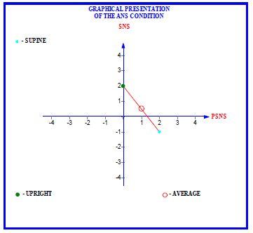
The Schellong test, in which blood pressure and pulse are measured lying down and standing, has long been known. The HRV measurement is modelled on this proven procedure and likewise measures the autonomic nervous system lying down and standing. This produces a picture of the parasympathetic and sympathetic systems.
The measurement requires a special device that transmits the electrical signals of the heart via a chest strap. The measurement is therefore completely harmless, like an ECG. It should be carried out on a normal day that reflects your everyday life well, and not when you are exceptionally stressed or acutely ill.
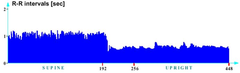
In a healthy person, the PNS is active when lying down and withdraws when standing; the SNS is active when standing and withdraws when lying down. Lying down, HRV measures the activity of the “Intrinsic Cardiac Nervous System”, a complex network of nerves at the heart that consists mainly of sympathetic and parasympathetic nerve cells. On standing up, the entire blood vessel system has to react and constrict the vessels slightly so that the blood does not sink into the legs. Standing up therefore tests not only the heart but also the sympathetic (and in part the parasympathetic) nerve supply to the smooth muscles of all blood vessels. In this way the test covers a large part of the nerve cells of the ANS and, by proxy, allows statements about its overall condition.
Two examples
What do I do with the result?
If the autonomic nervous system is weakened, the doctor can, together with further examinations, recommend individual treatments on this basis. With micronutrients tailored to your personal requirements, the ratio between phases of tension and recovery, and thus heart rate variability, can be favourably influenced.
Because the measurement is simple and inexpensive, it is excellently suited to documenting the success of therapy.
Homocysteine
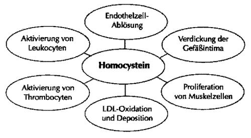
In ageing people, the most important chronic diseases are limited to a few underlying conditions. Of these, the arteriosclerotic changes that calcify the vessels come first. The fact that ever younger people are affected, however, also depends on our modern way of life. Risk factors: smoking, no natural, unprocessed foods with a lack of vitamins and trace elements, overweight, lack of exercise and stress. Here the much-cited cholesterol plays a subordinate role. Until now it has tended to distract from the more important causes of arteriosclerosis and has thus done the prognoses more harm than good. Thus raised cholesterol was found in only 35% of heart patients. The non-cardiovascular deaths among men and women have tended to increase with the reduction of cholesterol. Digestive disorders and tumours occurred in particular. In women there is in fact no scientific evidence at all that cholesterol actually plays any role in vascular damage, since the studies to date included only men. An uncritical transfer of the data to women is not legitimate. It is therefore time to seriously question the old dogma of the cholesterol hypothesis. In the search for other risk factors, homocysteine, a toxic amino acid, was first discovered at the end of the 1980s (amino acids are needed for protein production, in which homocysteine is an intermediate product). In the relatively rare enzyme deficiency diseases in which this amino acid accumulates, those affected die at a young age of pronounced arteriosclerosis and thromboses. This “homocysteinaemia” is, however, also encountered relatively frequently in a milder form. We find genetic conditions in 1 in 70 (!) people. Even more often the cause lies in a vitamin deficiency (folic acid, vitamin B6, B12, choline, riboflavin) due to poor nutrition, alcohol, smoking, in insufficient absorption (permeability) through the small intestine, in anorexia, radiation, chemotherapy or other medication. The latter are above all gastric acid blockers, anti-epileptic drugs, antidepressants, the contraceptive pill and others. It can be assumed that such serious, widespread chronic diseases as cataracts, osteoporosis, impaired brain performance, ageing skin, rheumatism etc. coincide with raised homocysteine levels. It is interesting that in people with Down syndrome heart attack, stroke or circulatory disorders are as good as unknown. Responsible for this is the surplus chromosome present here, which prevents homocysteine from rising.
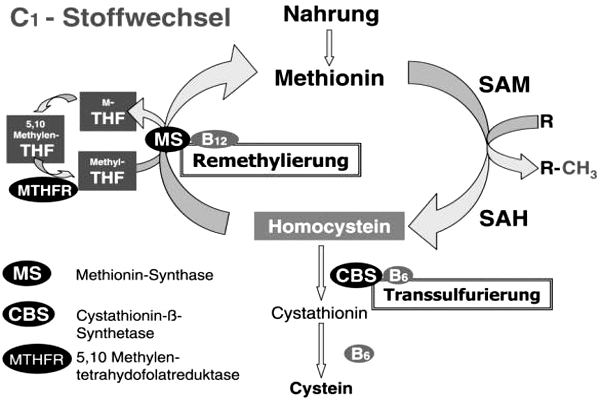
Biochemistry of homocysteine metabolism
Mechanism of arteriosclerosis
Homocysteine can be oxidised (become “rancid”) relatively easily, releasing hydrogen peroxide, which attacks the vessel walls directly and causes certain lipoproteins to become rancid as well. As homocysteine rises, blood clotting is promoted, so that the risk of thrombosis increases further. Only after homocysteine has caused primary damage to the vessel walls can cholesterol be deposited. Cholesterol therefore plays a subordinate role in the development of vascular diseases! People with kidney disease likewise suffer more often from vascular complications. This is explained by the fact that the kidneys are no longer able to excrete homocysteine. This also happens in diabetes.
Treatment
The precursor amino acids of homocysteine should be avoided as far as possible. These include methionine-rich foods such as meat. A vegetarian diet is low in methionine. You should therefore prefer foods rich in fibre, and avoid long storage times or overcooking food. Always wash fruit and vegetables whole and leave them in the water only briefly. Steam vegetables and use only a little liquid. Wherever possible, reuse the cooking liquid (e.g. for preparing sauces).
Make sure you get enough vitamin B6, B12 and folic acid. Unfortunately, however, the corresponding therapeutic doses can hardly be achieved with the usual diet.
Depending on the risk situation, these vitamins must additionally be taken or injected on an individual basis. In some circumstances supportive measures such as chelation therapy or haematogenous oxidation therapy would be advisable. However, a prior assessment is required in order to avoid unnecessary therapies.
Where homocysteine levels are clearly raised, a genetic examination of the “control enzyme” methylenetetrahydrofolate reductase MTHFR is indicated today (see fig. page 14: Homocysteine-Methyl-Transferase). A widespread polymorphism in the MTHFR gene leads to a variant of the MTHFR enzyme. The sequence variant has only about 50% of the activity of the wild-type enzyme. Individuals with this thermolabile form consequently have higher plasma levels of homocysteine. A base substitution in the coding region of the MTHFR gene is responsible. At position 677, the altered form shows a base substitution from C (cytosine) to T (thymine). In the amino acid sequence of the protein, this substitution results in a change from alanine to valine. The polymorphism is widespread. About 40% of the population are homozygous for the wild type (C677), 45% are heterozygous (C677 and T677) and 15% carry the sequence variant homozygously (T677). If a homozygous constellation has been demonstrated, a sufficient vitamin intake and corresponding nutritional measures, as stated above, must be ensured for life.
Lipid Exchange Therapy according to Patricia Kane
Lipid Exchange Therapy is used for two main indications
- Circulatory disorders
- Detoxification
CIRCULATORY DISORDERS
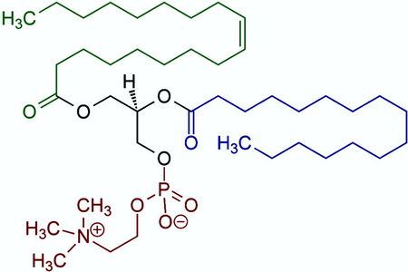
Lipid Exchange Therapy consists essentially of infusion treatments with essential phospholipids (EPL). The particularly effective component is phosphatidylcholine, which, when given intravenously, has been shown to be able to remove deposited fats from the inner vessel wall and, preventively, to stop cholesterol from being deposited again.
The main areas of application are promoting arterial blood flow in atherosclerosis, because it breaks down plaques, lowering the cholesterol level and dissolving fat emboli (globules of fat that block the vessels in the lungs).
Effects of Lipid Exchange
- reduces atherosclerotic plaque
- lowers LDL cholesterol
- raises HDL cholesterol
- helps with angina pectoris (tightness of the chest)
- extends walking distance in circulatory disorders of the legs
- improves mental functions
- improves sexual potency
Who needs Lipid Exchange Therapy?
In recent years our therapy centre has achieved very good results in the treatment of circulatory disorders - regardless of which parts of organs were affected. There is ample experience in the treatment of circulatory disorders of the coronary vessels, the vessels of the legs and the vessels of the brain. Associated complaints such as tightness of the chest, ringing in the ears (tinnitus), window-shopper's disease (claudication) and shortness of breath on exertion could be improved very well by Lipid Exchange infusions, without side effects. Walking distance becomes longer, the tightness of the chest disappears and impotence also responds well to the treatment.
The treatment is particularly suitable when the smallest vessels (capillaries) are affected, as for example in diabetes and in raised cholesterol levels (prophylaxis and therapy).
Lipid Exchange Therapy
The therapy consists of intravenous infusions, the number of which depends on the severity of the disease. The infusions are given on an outpatient basis, last 1.5 to 2 hours each and are not a strain. From experience we recommend combined treatment with Lipid Exchange and chelation infusions, alternating or in combination. This makes the treatment results better than with chelation infusions alone. The exact therapy plan is determined individually according to the initial medical findings. A basic treatment, during which treatment successes already become visible, consists of 20 infusion treatments on average - depending on the severity of the underlying disease.
The best therapy is a combination of Lipid Exchange and chelation therapy. After as few as 5 Lipid Exchange infusions the patient often notices a perceptible improvement in blood flow in the affected area. Medication can be reduced, and the patient is more capable again. Sexual potency is restored, walking distances become longer and the patient with angina pectoris has far fewer attacks.
Studies with laboratory animals also showed an extension of lifespan by 36% on average. An anti-ageing effect is therefore hoped for in humans as well.
DETOXIFICATION
Patricia Kane and Ed Kane have, in particular on the basis of fatty acid analysis as well, developed special protocols by which neurotoxins such as heavy metals, but also bacterial toxins and other toxic substances, can be removed above all from the predominantly fatty brain and nerve tissue and from the toxin-storing organs, liver and gall bladder, and by which an effective regenerative therapy of impaired liver and nerve cells is achieved at the same time.
Detoxification by means of phospholipids
The health of the cell membrane is equivalent to the health of the entire organism. The worst toxins are fat-soluble and accordingly cling to fatty acids. They lodge, in the truest sense of the word, in fatty surroundings and in doing so weaken and disrupt the functions of the cell. The probable result is early cell death (apoptosis). In general, normal mitosis provides for new cell growth in order to maintain the health of the body, which unfortunately does not apply, or applies only to a very limited extent, in the case of nerve cells.
Because of the affinity of toxins for fats, they can easily pass from the dead cells into the new cells in turn. In the state of health - when sufficient glutathione and vitamin C are present to bind these toxins before they can lodge in the newly formed cells - the body can keep these toxic substances under control.
Aim of detoxification
By exchanging damaged fats for healthy ones (PC, see below), the process of destruction is to be halted and affected cells are to regenerate. The important detoxification molecule glutathione, which is likewise used in Lipid Exchange, also serves this purpose.
Phosphatidylcholine (PC)
is the phospholipid most commonly found in the cell membrane. Because of its fatty acid content and because of the fat-soluble properties of toxins, fat-based therapies are also needed in order to be able to eliminate toxins.
Studies
Extensive research into the effect of PC has shown that it protects the liver from damage by alcohol, medicines, environmental toxins and foreign biological substances, as well as from infections by viruses, bacteria and fungi (Lieber 1994a, 1994b, 1995, 2001a, 2001b).
In the groundbreaking 1973 study by Wallnoefer and Hanusch, 650 patients with varying degrees of liver damage were observed for 5 years. The patients received 950 mg of PC intravenously and, in addition, 450-700 mg of PC orally. As soon as the blood values were normal, the patients received PC only orally. The patients were divided into the following categories according to the degree of their liver damage:
- -degenerated fatty liver,
- -acute inflammation,
- -chronic aggressive inflammation and
- -advanced cell destruction.
All categories benefited clearly from treatment with PC: in many patients the degenerated fatty liver could be cured completely, and in those with acute inflammation the healing process was accelerated by 10 days on average.
The use of anti-epileptic drugs frequently leads to liver poisoning. The status of patients who had received such drugs for 5 years on average was determined by means of the increase in their GGT value (Hisanaga 1980), and they were given PC for 6 months. This achieved a consistent improvement in the status of the patients and a lowering of their GGT values. Kuntz (1965) achieved similar improvements by giving PC to patients with chemical poisoning, and Esslinger (1966) in patients with plant poisoning.
Repeated viral attacks are a major challenge for the liver. In many controlled trials the administration of PC has proved to be a safe and effective measure against viral infections. (Mueting 1972, Hirayama 1980, Yamo 1978, Kosina 1981, Jenkins 1982, Visko 1985, Hantak 1990, Ilic and Begic-Janev 1991). In these studies optimal results were achieved when the patients were given PC both orally and intravenously in higher doses. As soon as the clinical values returned to normal, the patients received PC only orally.
These studies showed that treatment with PC reduced the liver enzymes, serum lipids, immune markers and bilirubin, and liver biopsies established that degenerated fatty livers, acute inflammations, jaundice, liver swellings and fibrosis were cured.
Patricia Kane had astonishing successes in patients with ALS or Parkinson's through the administration of PC together with glutathione. Patients confined to a wheelchair were again able to cover a few steps independently.
Main indications
- Liver detoxification
- Nervous disorders
Veins - sclerotherapy of varicose veins
The circulation of the legs is based on a complex interplay of the arteries, veins, lymph vessels and muscles. The oxygen-poor, used blood has to be conveyed back to the heart through the venous system against gravity. To make this task easier, the veins have valves which, like non-return valves, prevent the blood from flowing back. In this way the blood is literally pumped back through the veins that are compressed by the muscles when walking (“deep veins”) (the “peripheral heart” of the body). That is why walking is healthy and standing is bad.
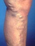
The superficial veins, which become apparent as varicose veins when they dilate, do not have this protective sheath of muscle. Although venous valves are found here too, they therefore tend to be widened by the weight of the blood lying above them. This prevents the valves from closing intact, so that the blood can flow back in the opposite direction. A vicious circle has arisen. In the end the blood flows downwards through the superficial veins instead of upwards. The skin is thus supplied with more oxygen-poor blood, which results in leg ulcers and poorly healing wounds. Likewise, the deep venous system has to cope not only with the normal blood but also with the blood flowing back, which in the long run can lead to overload (calf cramps, swelling, heavy legs).
Timely sclerotherapy or vein surgery can effectively prevent this and at the same time represents one of the most important medical indications for their treatment. The small varicose veins and the bothersome networks of spider veins, by contrast, are medically insignificant but occupy an important place cosmetically. While the large, thick varicose veins should rather be left to the surgeon, the small and medium-calibre ones are the domain of sclerotherapy (= sclerosing). Women suffer from varicose veins approx. twice as often as men.
Sclerotherapy of the veins
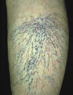
A sclerosing agent is introduced into the vein in order to produce an artificial inflammation of its walls. Some of these agents contain iodine, which is why we must be told of an iodine allergy without fail. The smaller veins are injected directly; from the large ones the blood is first drained. The bleeding stops once the leg is raised. The vein is now empty and collapsed. The sclerosing agent, now injected, can take effect at full concentration.
The compression bandage
After that, the leg has to be bandaged well so that the irritated vein walls are pressed together and can thus stick together. As a rule several bandages are needed for this. A small self-adhesive bandage over the sclerotherapy site must be left in place until the evening of the following day. Caution: above all near the ankle and knee joints, the skin under the bandage can become irritated and chafed (due to movement). Should pain occur, you must remove the bandage and take a look. The elastic bandages must be removed in the evening before going to sleep and put on tightly again in the morning. The technique of bandaging has to be learnt! Make sure you are instructed well. If the toes turn blue, this does not mean that the compression is too tight. Only when pain or cramps occur should the bandage be loosened. After removing the bandages and before putting them on, the vein gel prescribed for you must be rubbed in at the puncture site. In the course of the treatment, small lumps - clotted blood between the vein ends that have stuck together - may form at the sclerotherapy site. As a rule they disappear after months but, depending on their size and painfulness, can be punctured and squeezed out.
To achieve optimal results, the legs should be bandaged for at least four weeks after the last sclerotherapy injection.
The compression bandage is to be put on in the morning immediately after getting up. Do not walk around first, otherwise the decongestion of the leg that has taken place during the night is undone again. For effective decongestion the legs should, if possible, be elevated at night. However, the hollows of the knees must not sag in the process. So raise the bed at the foot end; a wedge pillow at the foot end is not enough.
- To put on the bandage, the foot, supported on the edge of a chair, must always be placed at a right angle
- Depending on the size and circumference of the leg, bandages 8 cm or 10 cm wide are best suited for compression bandages on the lower leg.
- Take the bandage in your hand so that the rolled-up part of the bandage lies on top and points outwards. Only in this way can the bandage be unrolled on the leg.
- When putting it on, unroll the bandage directly on the skin and pull both edges evenly in the direction of unrolling. Never pull away from the leg, otherwise the two edges of the bandage are tensioned unevenly and strangulating constriction furrows arise.
- The pressure exerted by the bandage must be strongest in the ankle region and decrease slowly towards the knee. Too much pressure in the calf area causes dangerous congestion. So pull the bandage tighter at the ankle and then ease off slowly, in keeping with the shape of the leg.
- You can check that the pressure is right by the fact that the toes first turn slightly bluish when the bandage is put on, but take on their natural skin colour again when walking.
- The foot area from the base joint of the toes and the heel are always bandaged, so that all congestion can only be pressed upwards.
- As a general rule, bandage dressings hold better and longer if you put a second bandage over the first one in the opposite direction of unrolling.
Sclerotherapy of varices is generally contraindicated in patients confined to bed, in arterial occlusive disease, deep vein thrombosis, diabetes mellitus, nephritis, active tuberculosis and other febrile or purulent infections, acute severe heart disease, all diseases and situations associated with restricted mobility (e.g. advanced age), and in all patients in whom compression therapy is contraindicated. Leg oedema is to be decongested by compression therapy before sclerosing.
SUMMARY
| Sclerotherapy | Operation |
|---|---|
| cheaper no hospitalisation / anaesthesia no time off work can / must as a rule be repeated at certain intervals (years) | good if afraid of injections better long-term results leaves scars |
| RISKS | |
| allergic reactions (rare) extensive inflammation of the veins permanent skin marks due to injection of the sclerosing agent next to the vein (hardly possible with correct technique) | deep vein thromboses (extremely rare) nerve injuries bruising injuries to main blood vessels infections |
Additional vein therapy
- Exercise! Walk longer distances (more than 7 steps!)
- Weight reduction
- Elevate the feet
- Avoid intensive exposure to sun and heat
- Shower the legs with cold water
- Compression treatment (support stockings etc.)
- Horse chestnut preparations to be taken orally