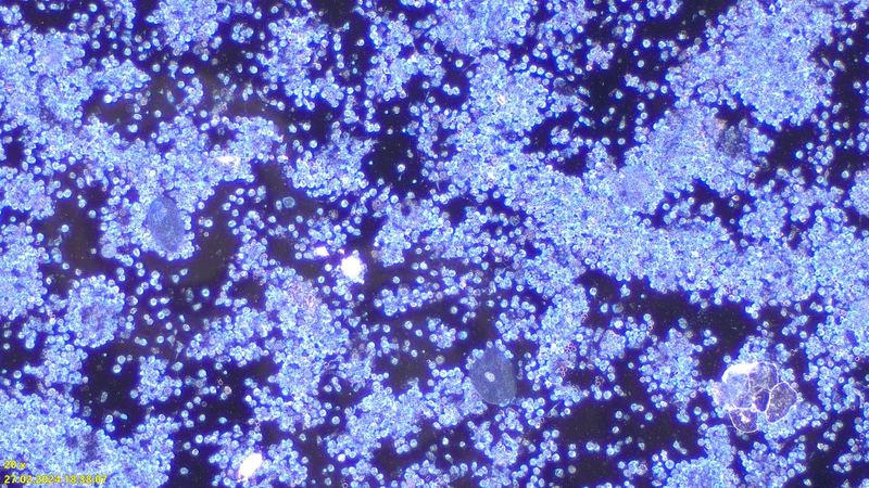
A special way of looking at the blood.
With this special examination, the living blood is observed directly under the microscope. With the usual methods, the specimen is first dried, finally stained and then, on top of that, assessed in ordinary transmitted-light microscopy, which has the same effect as when we try to look at something against the light. The fine structures are completely lost in the process. In the dark field, by contrast, the light is shone in from the side, and the details become visible.
These changes give us pointers regarding treatment. Special products, autologous blood therapy and infusions, which are worked out individually, serve this purpose.
Clear seawater: light from above
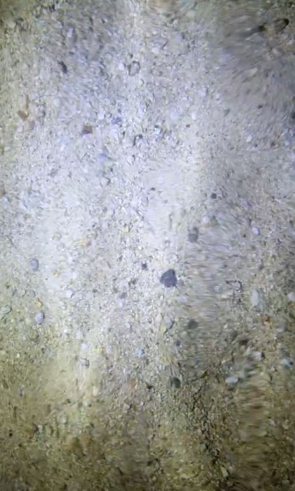
Seawater in the dark field: light from the side
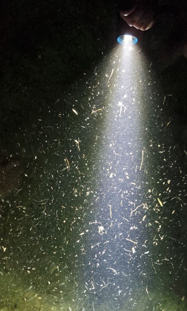
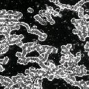
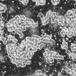
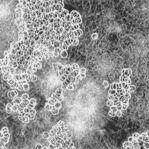
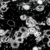
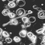
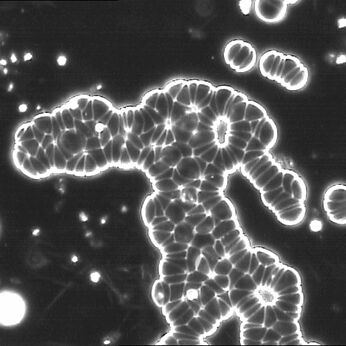
Just as here in the seawater example, considerably more detail will be seen in the dark field of the blood than in ordinary transmitted-light microscopy.
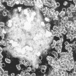
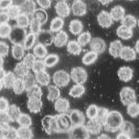
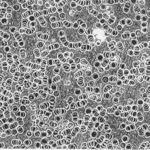
Dark-field examples of fresh blood
Background information
The primal property “life” is fundamentally immortal and embodies a tremendous power to preserve and unfold itself. Everyone carries this “immortality” within them. It does not die in a cremation and it survives the most hostile environments. It was thus possible to cultivate life forms even from hard coal, crude oil and even meteorites. Let us think of the dust of six-thousand-year-old mummies, which was still capable of triggering serious diseases (“curse of the pharaohs”). Discovering and describing this primal life is the achievement of Professor Günther Enderlein, who had recognised the possibilities of dark-field microscopy. It is no longer the cell that represents the smallest living unit, but the “protit”, tiny particles of approx. 1/100,000 mm in diameter with the incredible ability to germinate depending on the milieu of their surroundings. “Protit” was derived from the Greek Proteus, the uncanny god who could take on any shape and whose nature therefore could not be grasped. We also find these protits and the “most varied life forms” in our blood with dark-field microscopy, and depending on the composition of the blood these protits can take on other “shapes”, which may on the one hand be harmless and on the other hand be a burden on health. In the latter case one speaks of an “endobiosis”. For treatment it is important that, depending on lifestyle and therapy, this development can also be reversible!
All microbes accordingly pass through a species-specific cycle, such as textbook bacteriology accepts as a matter of course for malaria but to this day refuses to recognise for bacteria and fungi, although in the whole of nature there is no exception to the law of eternal change and of the unity of the macrocosm with the microcosm. Compare the maxim of the famous ancient Chinese physician Lao Tse: “When things have reached their full unfolding, each returns to its origin.”
What does conventional medicine say about this? After all, the various germs are well known! It was the assertion of the famous researcher L. Pasteur that all microbes are unchangeable, that each species produces only one specific disease, that bacteria and fungi never arise through spontaneous generation, and that blood and tissue are sterile in the healthy state. Diseases, he held, have their origin in the bacteria, which attack the body from outside and descend from pre-existing bacteria. The scientist Claude Bernard fought against this and corrected: “No, gentlemen, the microbe is nothing. The breeding ground is everything.” Only on his deathbed is Pasteur said to have admitted: “Bernard was right after all...”. In the meantime, however, medical thinking had become so entrenched in Pasteur’s philosophy that a rethink was no longer possible.
2 macrophages at the bottom left attack a foreign body, time lapse
The two white blood cells know the way and push the red blood cells aside in order to get to the “enemy”. Once again one can only marvel at the power and superiority of nature and, in this case, of our immune system.
Dark-field image from the SGK laboratory.
Urine sediment in the dark field
A further milestone in the assessment of kidney diseases is the dark-field analysis of the urine sediment. The resolution is many times that of so-called transmitted-light microscopy. In addition, in conventional microscopy the specimen has to be dried and stained for anything to be seen at all. The cells die in the process. The specimen is dead. Dark-field microscopy makes it possible for the first time to analyse the living urine. You can see how exciting and revealing that can be in the video below.
Masses of white blood cells in inflammation